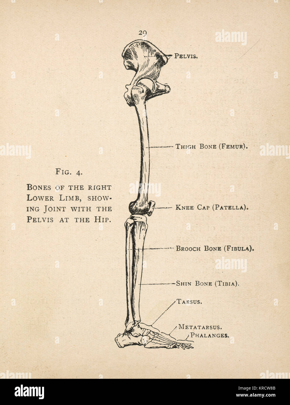
Leg Bones Diagram : Leg Anatomy. Your leg bones are very large and strong to help support the weight of your body. Skeleton leg ankle joints and toe phalanges, cuboid, metatarsal, navicular and cuneiform bones, hand drawn dorsal view of foot. The bones involved in it, however, are only the femur and the tibia, although the smaller bone of the leg, the fibula, is carried along in the movements of flexion, extension, and slight rotation that this joint. The human leg consists of 8 bones, 4 per leg. Human foot bones anatomy sketch of orthopedics medicine.
High quality realistic skeleton legs. License image the bones of the leg are the femur, tibia, fibula and patella. Learn vocabulary, terms and more with flashcards, games and other study tools. The bone that goes from your pelvis to your knee is called the femur (say: Visit kenhub for more skeletal system quizzes.
Ballet Feet | ARHtistic License from arhtisticlicense.files.wordpress.com Bone surfaces at synovial joints are protected by a coating of articular cartilage. Time to jump right into the biggest and strongest bones in the human body. The bones involved in it, however, are only the femur and the tibia, although the smaller bone of the leg, the fibula, is carried along in the movements of flexion, extension, and slight rotation that this joint. High resolution textures and displacement included. Synovial joints are often supported and reinforced by surrounding ligaments, which limit movement to prevent injury. At the same time, the bones and joints of the leg and foot must be strong enough to support the body's weight while remaining flexible enough for movement and balance. Bones of the leg and foot, lower leg bone anatomy, leg bones anatomy, leg muscles, leg bones diagram, leg bone structure, leg anatomy muscles, parts of the lower leg. Interactive anatomical atlas of the head, brain, and neck based on anatomical diagrams and ct and mri medical imaging exams.
Human foot bones anatomy sketch of orthopedics medicine.
He leg's main function in the human is for locomotion and support of the rest of the body. Diagram and names of leg bones, diagram of foot and leg bones, diagram of leg bones, diagram of lower leg related posts of diagram of leg bones. Bones of the leg and foot, lower leg bone anatomy, leg bones anatomy, leg muscles, leg bones diagram, leg bone structure, leg anatomy muscles, parts of the lower leg. High quality realistic skeleton legs. Human foot bones anatomy sketch of orthopedics medicine. The bone that goes from your pelvis to your knee is called the femur (say: Most bones (particularly the long bones of the arms and legs — which make up the appendicular skeleton) have a hard outer shell known as cortical bone. The bones and joints in the feet experience wear and tear, so conditions that cause damage to the it is usually the result of a muscle imbalance when the long muscles of the lower leg overpower the. Learn how to draw the femur, patella, tibia, and fibula in this lesson! Time to jump right into the biggest and strongest bones in the human body. Synovial joints are often supported and reinforced by surrounding ligaments, which limit movement to prevent injury. The bones involved in it, however, are only the femur and the tibia, although the smaller bone of the leg, the fibula, is carried along in the movements of flexion, extension, and slight rotation that this joint. Your leg bones are very large and strong to help support the weight of your body.
The bones involved in it, however, are only the femur and the tibia, although the smaller bone of the leg, the fibula, is carried along in the movements of flexion, extension, and slight rotation that this joint. The bones of the leg are the femur, tibia, fibula and patella. However, the definition in human anatomy refers only to the section of the lower limb extending from the knee to. He leg's main function in the human is for locomotion and support of the rest of the body. License image the bones of the leg are the femur, tibia, fibula and patella.
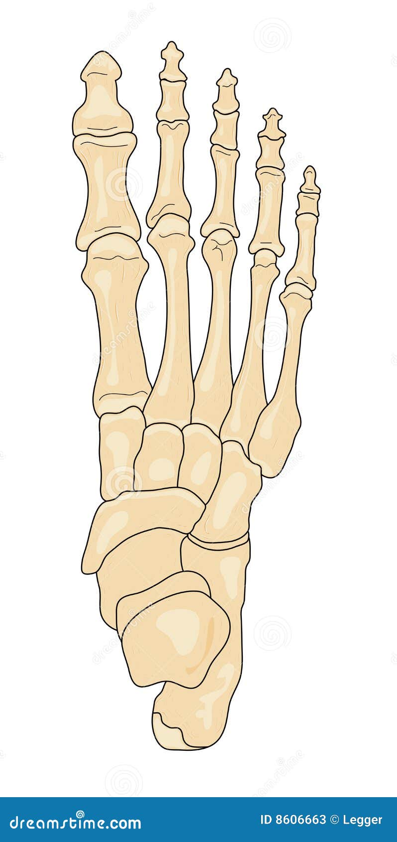
Bones of the Foot stock vector. Illustration of ankle - 8606663 from thumbs.dreamstime.com This bright worksheet helps your child bring these technical terms down to size. Your legs are two of your most important body parts. A leg bone is a bone found in the leg. Bones of the leg and foot, lower leg bone anatomy, leg bones anatomy, leg muscles, leg bones diagram, leg bone structure, leg anatomy muscles, parts of the lower leg. Learn how to draw the femur, patella, tibia, and fibula in this lesson! The foot bones shown in this diagram are the talus, navicular, cuneiform, cuboid, metatarsals and calcaneus. Interactive anatomical atlas of the head, brain, and neck based on anatomical diagrams and ct and mri medical imaging exams. High resolution textures and displacement included.
The foot bones shown in this diagram are the talus, navicular, cuneiform, cuboid, metatarsals.
The foot bones shown in this diagram are the talus, navicular, cuneiform, cuboid. Synovial joints are often supported and reinforced by surrounding ligaments, which limit movement to prevent injury. Use the leg bones diagrams to learn the names of the leg bones. Bones of the leg and foot, lower leg bone anatomy, leg bones anatomy, leg muscles, leg bones diagram, leg bone structure, leg anatomy muscles, parts of the lower leg. Diagram and names of leg bones, diagram of foot and leg bones, diagram of leg bones, diagram of lower leg related posts of diagram of leg bones. Human foot bones anatomy sketch of orthopedics medicine. These simple labelled diagrams of the bones of the lower legs and feet and the bones of the arms and hands this diagram shows the skeletal structure of the leg (anterior view) and foot (dorsal view). He leg's main function in the human is for locomotion and support of the rest of the body. Bone surfaces at synovial joints are protected by a coating of articular cartilage. The human leg consists of 8 bones, 4 per leg. High resolution textures and displacement included. Start learning with our skeleton diagrams, bone labeling exercises and skeletal system quizzes! The bone that goes from your pelvis to your knee is called the femur (say:
Time to jump right into the biggest and strongest bones in the human body. These can include any the following: This bright worksheet helps your child bring these technical terms down to size. Your legs are two of your most important body parts. Learn the bones of the body with skeletal system quizzes.
Human Foot Anatomy Stock Photos & Human Foot Anatomy Stock Images - Alamy from c8.alamy.com Diagram and names of leg bones, diagram of foot and leg bones, diagram of leg bones, diagram of lower leg related posts of diagram of leg bones. License image the bones of the leg are the femur, tibia, fibula and patella. Use the leg bones diagrams to learn the names of the leg bones. The foot bones shown in this diagram are the talus, navicular, cuneiform, cuboid, metatarsals and calcaneus. At the microscopic level, this hard outer. Learn the bones of the body with skeletal system quizzes. Skeleton leg ankle joints and toe phalanges, cuboid, metatarsal, navicular and cuneiform bones, hand drawn dorsal view of foot. At the same time, the bones and joints of the leg and foot must be strong enough to support the body's weight while remaining flexible enough for movement and balance.
Interactive anatomical atlas of the head, brain, and neck based on anatomical diagrams and ct and mri medical imaging exams.
High quality realistic skeleton legs. High resolution textures and displacement included. At the microscopic level, this hard outer. The bones involved in it, however, are only the femur and the tibia, although the smaller bone of the leg, the fibula, is carried along in the movements of flexion, extension, and slight rotation that this joint. Bone surfaces at synovial joints are protected by a coating of articular cartilage. The foot bones shown in this diagram are the talus, navicular, cuneiform, cuboid. Learn how to draw the femur, patella, tibia, and fibula in this lesson! Synovial joints are often supported and reinforced by surrounding ligaments, which limit movement to prevent injury. Your leg bones are very large and strong to help support the weight of your body. Visit kenhub for more skeletal system quizzes. The foot bones shown in this diagram are the talus, navicular, cuneiform, cuboid, metatarsals and calcaneus. These can include any the following: He'll boost his body knowledge as he matches up the names of the bones with their proper places on the leg diagram.
Cute Boys Speedo Bulges - Pin on Real Wrestlers . Wonder how many days of juice is stored in that manhood. Find a range of boys speedo swimwear available here. See, that's what the app is perfect for. Submitted 1 year ago by hungjack1999. As a boylover the dream is to be friends with boys and be able to watch ov. By jra on nov 6, 2019. It is much easier than making a girls swimsuit as there are less steps. Grey speedo boy is a classic. I like to see other big men do the same. See, that's what the app is perfect for. Bulge speedo cute hot gay straight men's swim | Gay on - Good Lookin - Underwear | Pinterest ... from s-media-cache-ak0.pinimg.com Gay, male, guys, speedos on men, speedos on boys, speedos on twinks, cute, hot, shirtless, smooth, pecks, abs, fitness, sports, speedo, tumblr, eye candy speedo bulge | tumblr. It is much easier t...
Para Jailbreak : Adjust Jailbreak In A Week Winter Mode For Apple Watch Floatytab Prysm . Direct download link for the latest version of evasion7: Ios 8.4.1, the latest ios software update cannot be. Cydia download ios 14.7 is the most trending topic these days because apple just released ios 14.7 for public use. Download the checkra1n jailbreak tool according to your operating system. There is no online method or mobile based method to install checkra1n yet. This page is the ultimate resource for every ios firmware available, download links for jailbreak tools such as, evasi0n, absinthe, redsn0w, etc, as well as links to some of our favorite softwares. Connect your usb flash drive to windows. Also you can find here all the valid jailbreak. Pangu jailbreak is a freeware ios jailbreak software download filed under iphone tools and made available by pangu team for windows. Checkra1n jailbreak has been released from ios 12 up to ios 14.7.1 jailbreak and available for ma...
Juego Mesa Serpiente - Juego 2 En 1 Serpientes Escaleras Juego Oca Mesa Casa | Mercado Libre . Aprende a jugar a la joya de la serpiente. Cocodrilos y serpientes juego de mesa los juegos del caracol. Juego de serpientes y escaleras, gouache sobre lienzo (india, siglo xix). Set multi juegos 10 en 1 clásicos. El juego es un concurso de carreras simple basado en pura suerte, y es popular entre los niños pequeños. El tesoro de la serpiente next point 1350 delmy. 8,49 € (2 nuevas ofertas) edades: Check spelling or type a new query. Juegos de mesa para niños juegos didacticos juegos para niños actividades para primaria proyectos escolares juegos de culebras serpientes y escaleras juego juegos de parques juegos de matemáticas preescolares. Juegos de mesa cocodrilos y serpientes o la oca tableros. Serpientes y Escaleras - Juegos de Mesa Clásicos for Android - APK Download from image.winudf.com ...


Comments
Post a Comment